Iridology, also known as iridodiagnosis or iridiagnosis, is an alternative medicine technique that assesses health conditions by examining the patterns, colors, and other characteristics of the iris. This practice posits that these elements reflect systemic health and provide insights into a patient’s wellness. It’s a non-invasive analysis method that can potentially identify the early stages of health concerns.
The history of iridology traces back to at least 3,000 BCE. Explicit principles of iridology were described in Philippus Meyeus’ 1665 work “Chiromatica Medica.”
The term “Augendiagnostik” (“eye diagnosis”, loosely translated as iridology) was coined by Ignaz von Peczely in the 19th century. Nils Liljequist’s observations and atlas in 1893 significantly contributed to the field of iridology. Pastor Emanuel Felke furthered iridological research in Germany in the early 1900s, establishing the Felke Institute, the leading center of iridological research and training.
In the 1950s, Bernard Jensen popularized iridology in the United States, emphasizing toxin exposure and natural detoxification. However, a 1979 attempt to establish diagnostic grounds faced challenges, highlighting the complexities of iris analysis in medical diagnosis.
Iridology, as a holistic health assessment tool, offers several advantages.
Firstly, it’s non-invasive, ensuring analysis without patient discomfort or risk. This makes it appealing for those averse to more intrusive procedures.
Secondly, iridology’s potential for early detection allows the identification of systemic imbalances before symptoms manifest, enabling preventive measures.
Lastly, its holistic perspective views the body as an interconnected system, offering comprehensive insights into overall health and facilitating the development of balanced, personalized plans.
Common findings that holistic iridology can reveal are explained below.
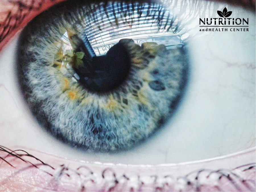
- Inflammation: Iridology helps identify signs of inflammation in the body. This is often represented by white or light-colored areas in the iris. The severity of inflammation can be gauged by the intensity of the white coloration.
- Toxicity: Iridology is used to assess toxic overload in the body. Dark or cloudy spots in the iris are thought to be indicators of toxic buildup.
- Predisposition to Certain Diseases: Iridology is often used to assess an individual’s susceptibility to certain health conditions. Irregularities in specific iris zones can reveal potential weaknesses in corresponding organs or systems of the body.
- Liver Damage: Iridology is used to assess liver health. A yellowish-brown discoloration or a cloudy appearance in the area of the iris associated with the liver might suggest liver-related issues.
Below are the common methods that iridologists use.
- Flashlight and Magnifying Glass: Iridologists commonly utilize a flashlight and magnifying glass during examinations to closely inspect the irises for tissue changes and specific features. This allows to observe intricate details, pigment patterns, and irregular stromal architecture.
- Cameras or Slit-Lamp Microscopes: Sophisticated equipment, such as cameras or slit-lamp microscopes, may be employed by iridologists for a more in-depth analysis of the irises. These tools enable a closer examination of subtle markings and patterns that could be indicative of underlying health conditions.
- Analysis of Pigment Patterns: Iridologists pay close attention to pigment patterns in the iris, as variations in color and texture can reflect changes in the corresponding body organs. Specific pigment patterns are examined to draw connections between the observed features and potential health conditions.
- Iris Chart Comparison: Iridologists refer to iris charts, which divide the iris into approximately 80–90 zones, correlating each zone with specific parts of the body. The markings and patterns observed during an examination are compared to these charts to identify potential associations with various organs and systems.